0260
High temporo-spatial resolution VASO reveals differential laminar reactivity to event-related stimuli at 7T1Faculty of Psychology and Neuroscience, Maastricht University, Maastricht, NL, Maastricht, Netherlands
Synopsis
Due to its high specificity, VASO plays a major role in laminar fMRI. To mitigate its lower sensitivity compared to GE-BOLD researchers mostly employ block-designs to increase SNR. Here, we developed a VASO sequence with a short TR (895ms volume acquisition) and showed that it provides the means to capture layer-specific haemodynamic responses with high spatio-temporal resolution. During event-related stimulation, we show reliable responses in visual and somatosensory cortices. Furthermore, the short TR and high specificity of VASO enabled us to show differences in laminar reactivity and onset times, thus demonstrating the high value of event-related designs using CBV-based fMRI.
Introduction
FMRI at ultra high resolutions lends the possibility to investigate the human brain at the mesoscopic scale (Dumoulin, 2016). A key feature at this level is the organization of cortical layers. While the commonly used GE-BOLD is biased towards the cortical surface, non-BOLD contrasts like Vascular Space Occupancy (VASO) have a higher specificity to the underlying microvasculature (Huber, 2017). However, this comes at the cost of lower sensitivity. Nevertheless, researchers have successfully used experimental conditions with long blocks of stimulation (~30s, e.g. Huber, 2017, but also see Persichetti, 2020). The possibility to employ short paced stimulation designs would widen the range of neuroscientific insights to be gained.The goal of the current study was:- To develop and test a VASO sequence protocol with a short TR which provides the means to capture layer-specific haemodynamic responses with high temporal resolution (895 ms volume acquisition TRs).
- To characterise the sub-millimeter detection sensitivity of block and event-wise stimulation.
- To investigate the generalizability of different stimulus modalities (vision and somatosensation).
Methods
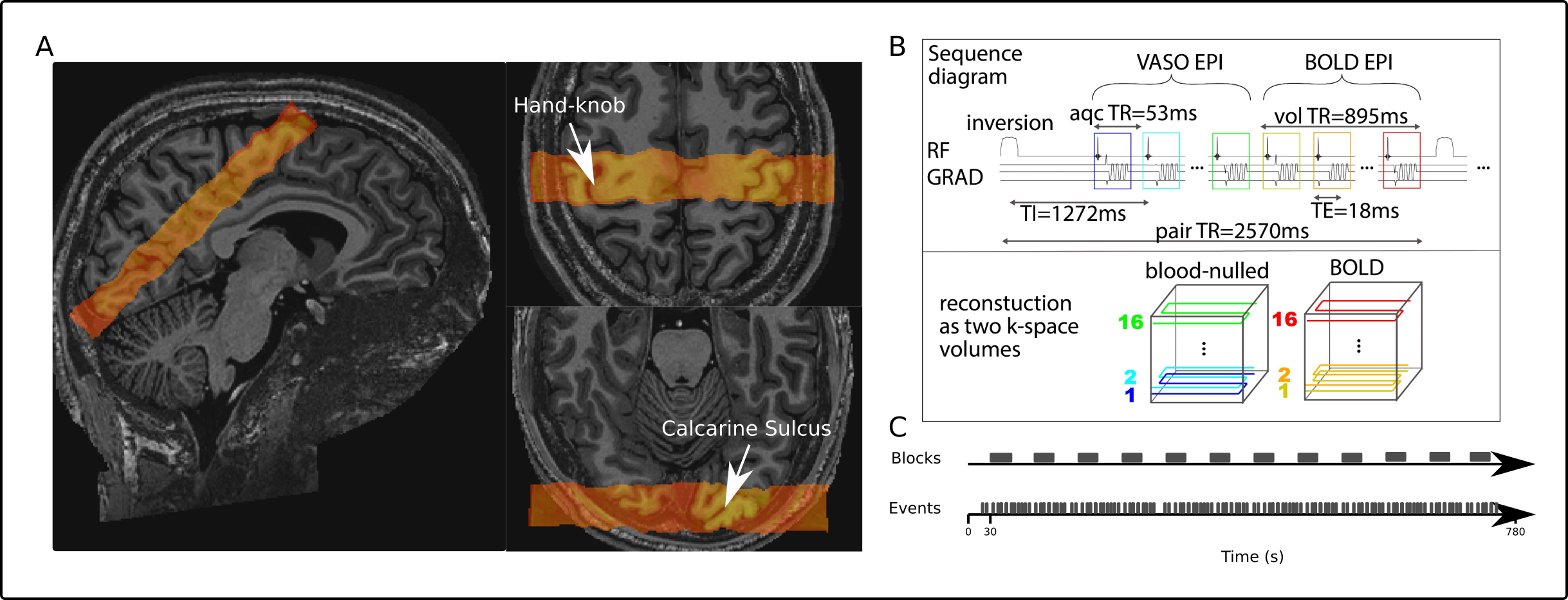
Data-acquisition: 4 participants underwent scanning using a ‘classical’ MAGNETOM 7T scanner (Siemens Healthineers, Erlangen, Germany), equipped with a 32-channel head coil at Scannexus (Maastricht, The Netherlands). All functional scans were performed with slice-selective slab-inversion VASO (SS-SI VASO, Huber, 2014) and a nominal resolution of 0.9mm isotropic (16 slices TI1/TI2/TR/TE = 50/825/1720/18 ms, partial fourier factor = 6/8, flip angle = 26°, FLASH GRAPPA 3, bandwidth =1266Hz/Px, FoV = 133 mm, Fig1 B). Slice position and orientation were chosen individually for each subject and covered the calcarine sulci and as much of the right postcentral gyrus as possible (Fig1 A). Finally, whole brain anatomical images (0.7mm isotropic) were acquired using MP2RAGE (Marques. 2010).Paradigm: During block-stimulation runs (2 runs of ~12 minutes), flickering checkerboards were presented at 16Hz and, concurrently, 5 digit-tips (left hand) were stimulated (30s on-off block design) by means of a piezoelectric vibrotactile stimulator (mini PTS system, Dancer Design, UK). During subsequent event-stimulation runs (2-4 runs of ~12 minutes), we presented the same visio-tactile stimuli for 2s per trial with inter-trial intervals (ITIs) between 3 and 10s chosen from a uniform distribution, resulting in 84 trials per run. These highly irregular ITIs were chosen to accurately capture the haemodynamic responses of BOLD and VASO. The stimulation pattern was generated once and then used for all runs in order to allow averaging.
Data-processing: Motion- and BOLD-correction was applied using ants-Registration and LAYNII, respectively. GLM-analyses were performed in FSL (contrast: stimulation vs rest), ROIs were manually drawn in FSLeyes and layering was performed on spatially upsampled data (factor 5 in-plane) using LAYNII (Huber, 2021). Finally, the ROIs were used to generate event-related averages (ERAs) by extracting timecourses from 4 TRs before and 8 TRs after stimulation and averaging them using custom python-scripts. For analysis code, see: https://bit.ly/3qdZCBG.
Results and Discussion
Here, we discuss the results in visual cortices. For blocks, z-maps show clear responses in all participants (Fig2 A). ERAs for both VASO and BOLD show the expected sustained response which is stronger for BOLD than for VASO (Fig2 B). Z-scores increase strongly towards the cortical surface for BOLD but less for VASO (Fig2 C). For unimodal responses to long stimuli, a peak in middle layers would be expected for VASO (under the assumption that the nulled blood has been drained between volume TRs). Possibly, the signal increase we see towards the surface for VASO and BOLD reflects the multimodal input from tactile stimulation. When averaging multiple runs, we also obtained reliable z-maps for event-related stimuli (Fig3 A) and ERAs that follow the expected temporal pattern, with clear peaks, initial dips and undershoots (Fig3 B). Importantly, the general response pattern stabilizes after averaging about 20 event-trials (Fig4 B), showing its suitability for the use in neuroscientifically valuable designs due to high efficiency. Still, the event-related results show a reliable, but lower detection sensitivity compared to the block design data (as seen by the less pronounced maps in Fig3 A vs. Fig2 A, lower signal change Fig3 B vs. Fig2 B and lower z-scores in Fig4 C). Importantly, Fig3 D shows earlier responses for middle and deep layers in VASO, which demonstrates its high value for depth-dependent analyses. The slow venous hemodynamic filter in BOLD, however, cannot capture these differences in our data.Similar results were obtained in the somatosensory cortex (S1, see Fig5). Note, that here we found higher z-scores for event-wise stimulation in VASO (Fig5 D).
Conclusion and Summary
Here, we characterize the applicability of SS-SI VASO with event-related stimulation. With a short 895 ms volume acquisition TRs, we were able to capture the haemodynamic response for VASO and BOLD within a few average trials. Furthermore, the short TR and high specificity of VASO enabled us to show subtle laminar-dependent timing differences of neural processing and vascular reactivity features. Here, deep and middle layers showed the earliest responses in VASO but not BOLD. We believe that this shows the added value of employing shorter stimulation periods using VASO, which greatly increases the range of possible design choices for CBV-based laminar fMRI.Acknowledgements
Laurentius Huber was funded by from the NWO VENI project 016.Veni.198.032.
Sebastian Dresbach is supported by the ‘Robin Hood’ fund of the Faculty of Psychology and Neuroscience and the department of Cognitive Neuroscience
Scanning was supported by FPN (faculty of psychology and neuroscience) via the MBIC grant scheme. Scanning was performed at the facilities of Scannexus B.V. (Maastricht, Netherlands).
References
- Dumoulin, S. O., Fracasso, A., van der Zwaag, W., Siero, J. C. W., & Petridou, N. (2018). Ultra-high field MRI: Advancing systems neuroscience towards mesoscopic human brain function. NeuroImage, 168(September 2016), 345–357. https://doi.org/10.1016/j.neuroimage.2017.01.028
- Huber, L., Ivanov, D., Krieger, S. N., Streicher, M. N., Mildner, T., Poser, B. A., … Turner, R. (2014). Slab-selective, BOLD-corrected VASO at 7 tesla provides measures of cerebral blood volume reactivity with high signal-to-noise ratio. Magnetic Resonance in Medicine, 72(1), 137–148. https://doi.org/10.1002/mrm.24916
- Huber, L., Handwerker, D. A., Jangraw, D. C., Chen, G., Hall, A., Stüber, C., … Bandettini, P. A. (2017). High-Resolution CBV-fMRI Allows Mapping of Laminar Activity and Connectivity of Cortical Input and Output in Human M1. Neuron, 96(6), 1253-1263.e7. https://doi.org/10.1016/j.neuron.2017.11.005
- Huber, L. (Renzo), Poser, B. A., Bandettini, P. A., Arora, K., Wagstyl, K., Cho, S., … Gulban, O. F. (2021). LayNii: A software suite for layer-fMRI. NeuroImage, 237(April), 118091. https://doi.org/10.1016/j.neuroimage.2021.118091
- Marques, J. P., Kober, T., Krueger, G., van der Zwaag, W., Van de Moortele, P. F., & Gruetter, R. (2010). MP2RAGE, a self bias-field corrected sequence for improved segmentation and T1-mapping at high field. NeuroImage, 49(2), 1271–1281. https://doi.org/10.1016/j.neuroimage.2009.10.002
- Persichetti, A. S., Avery, J. A., Huber, L., Merriam, E. P., & Martin, A. (2020). Layer-Specific Contributions to Imagined and Executed Hand Movements in Human Primary Motor Cortex. Current Biology, 1–5. https://doi.org/10.1016/j.cub.2020.02.046
Figures